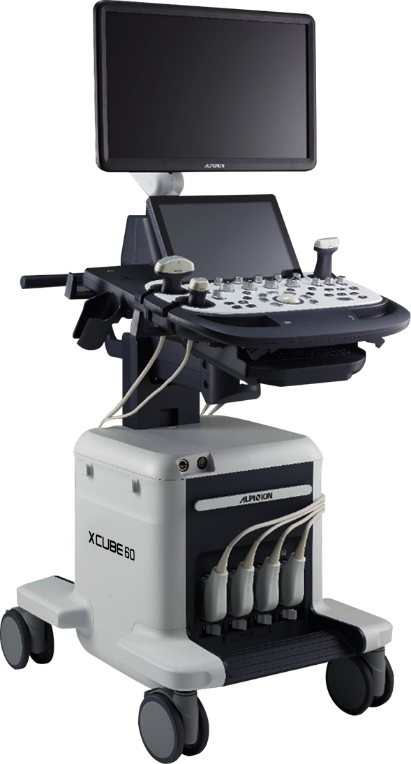
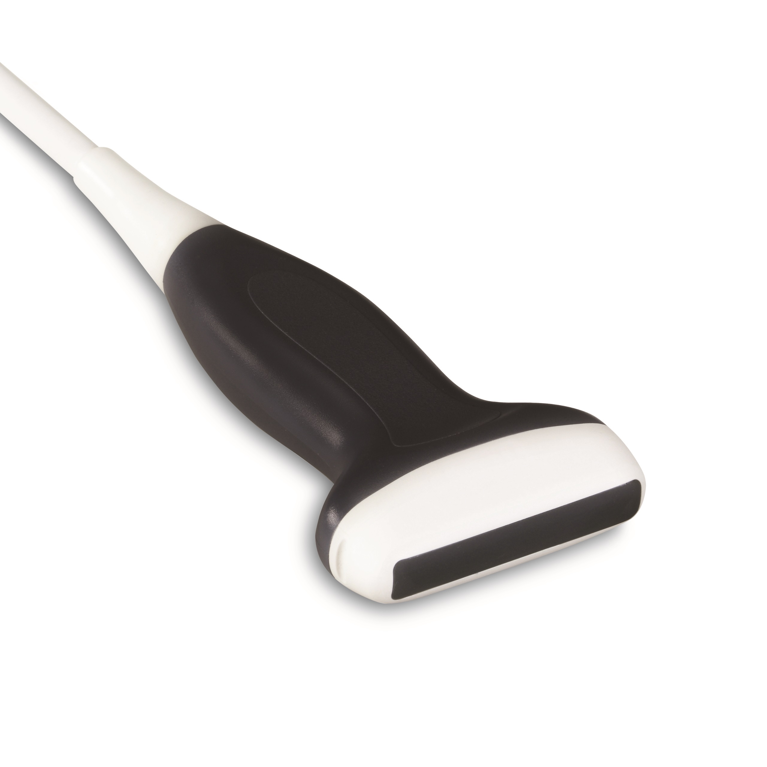
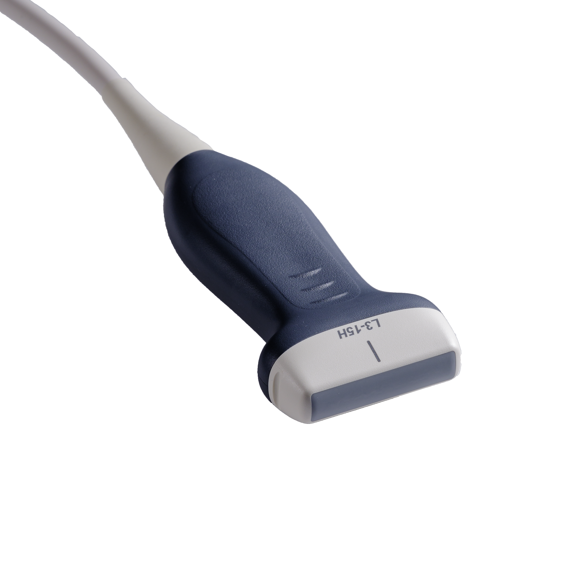
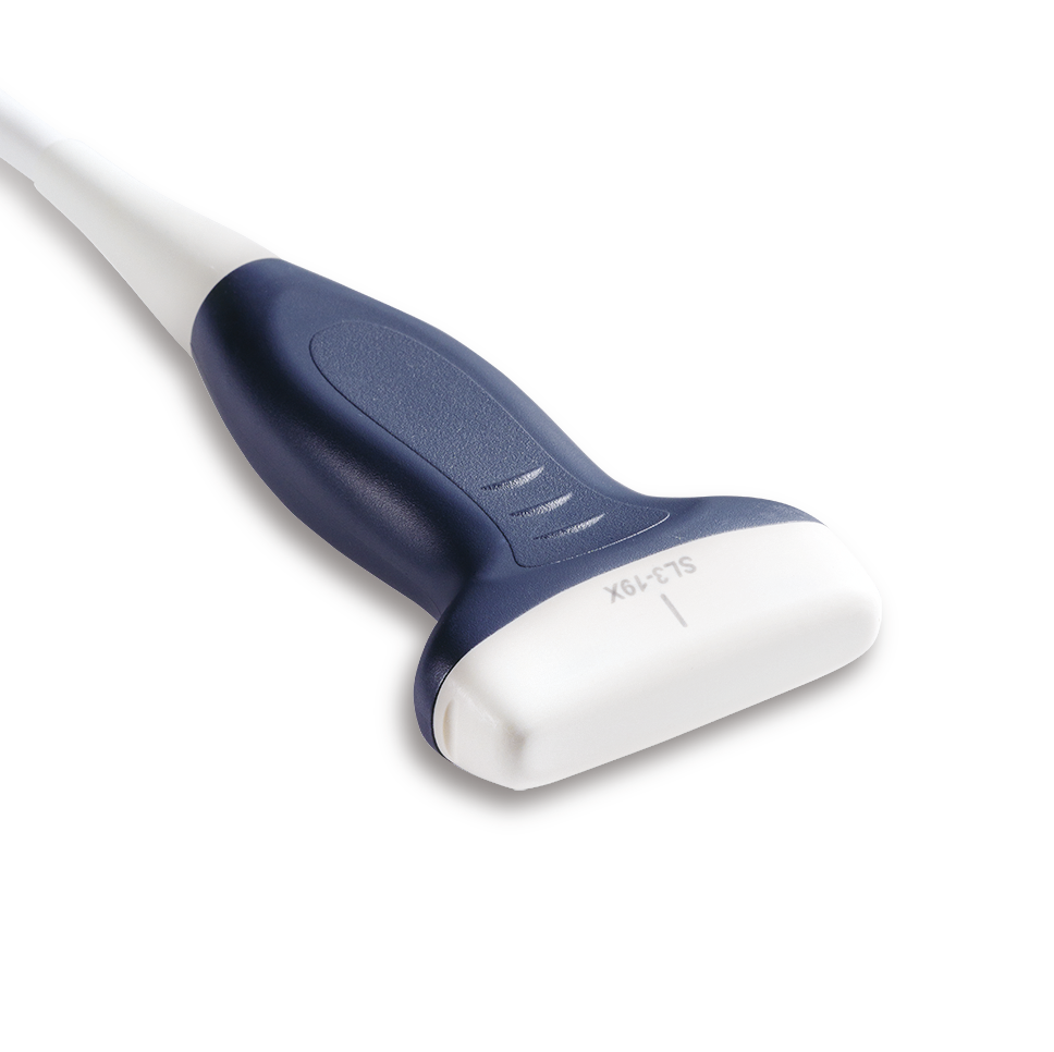
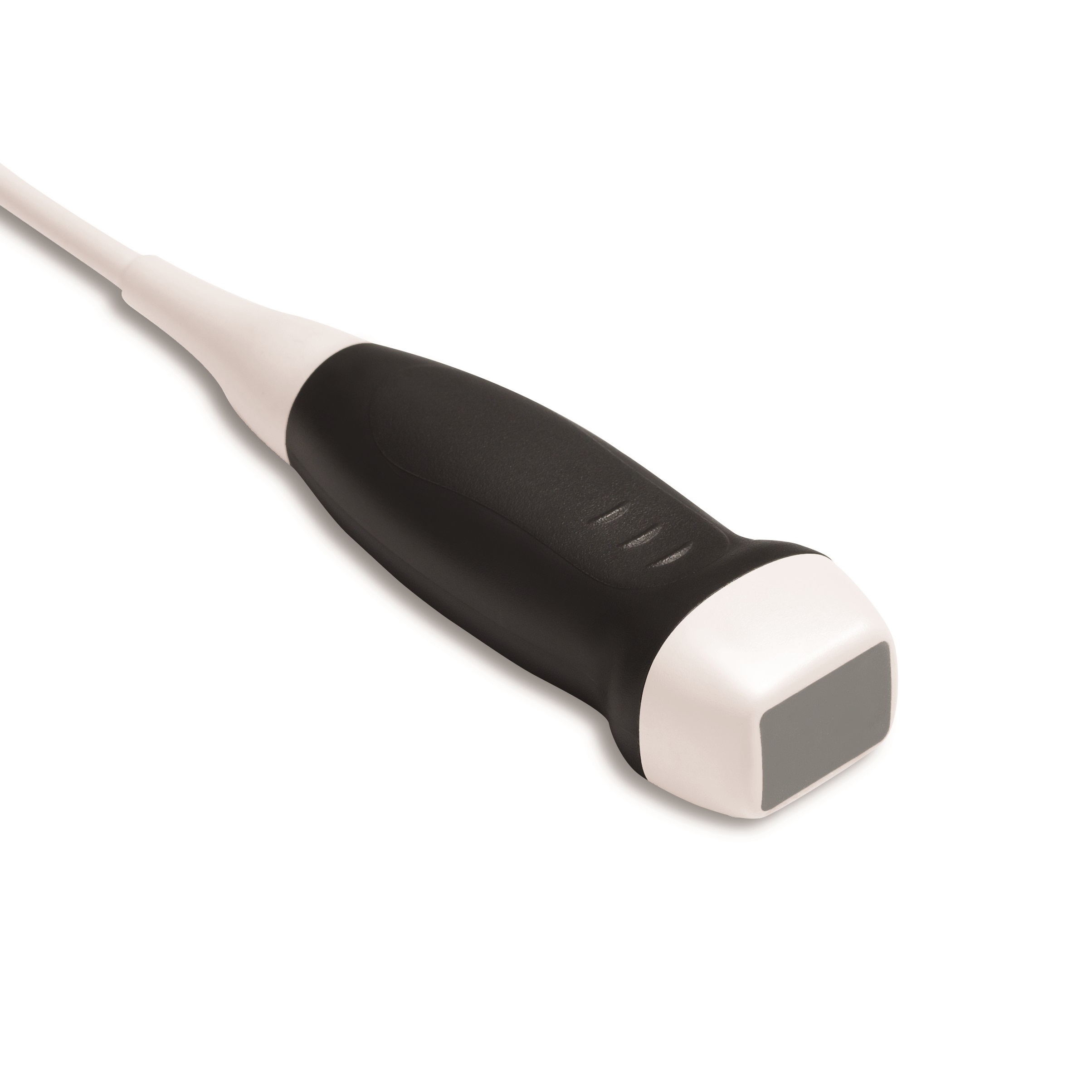
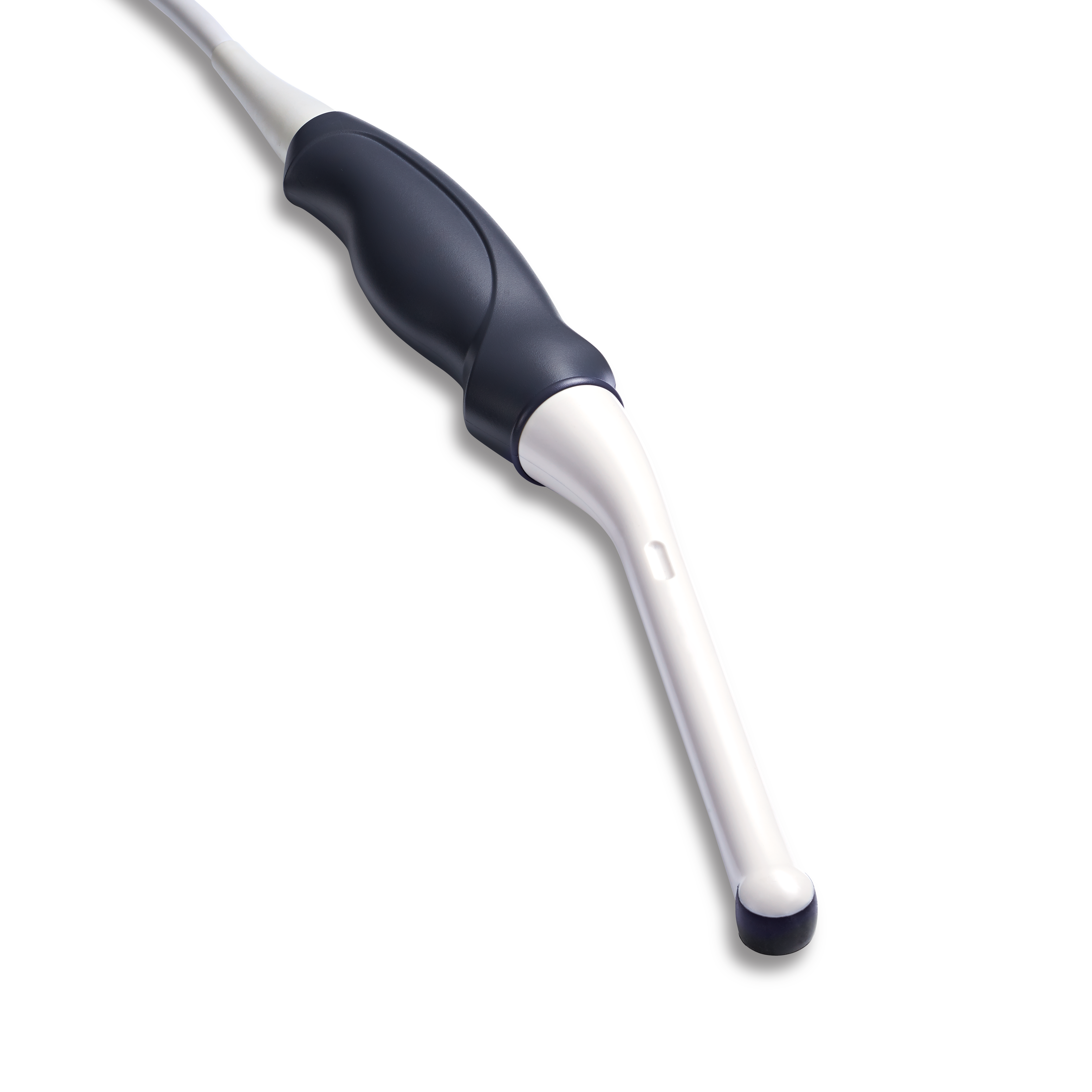
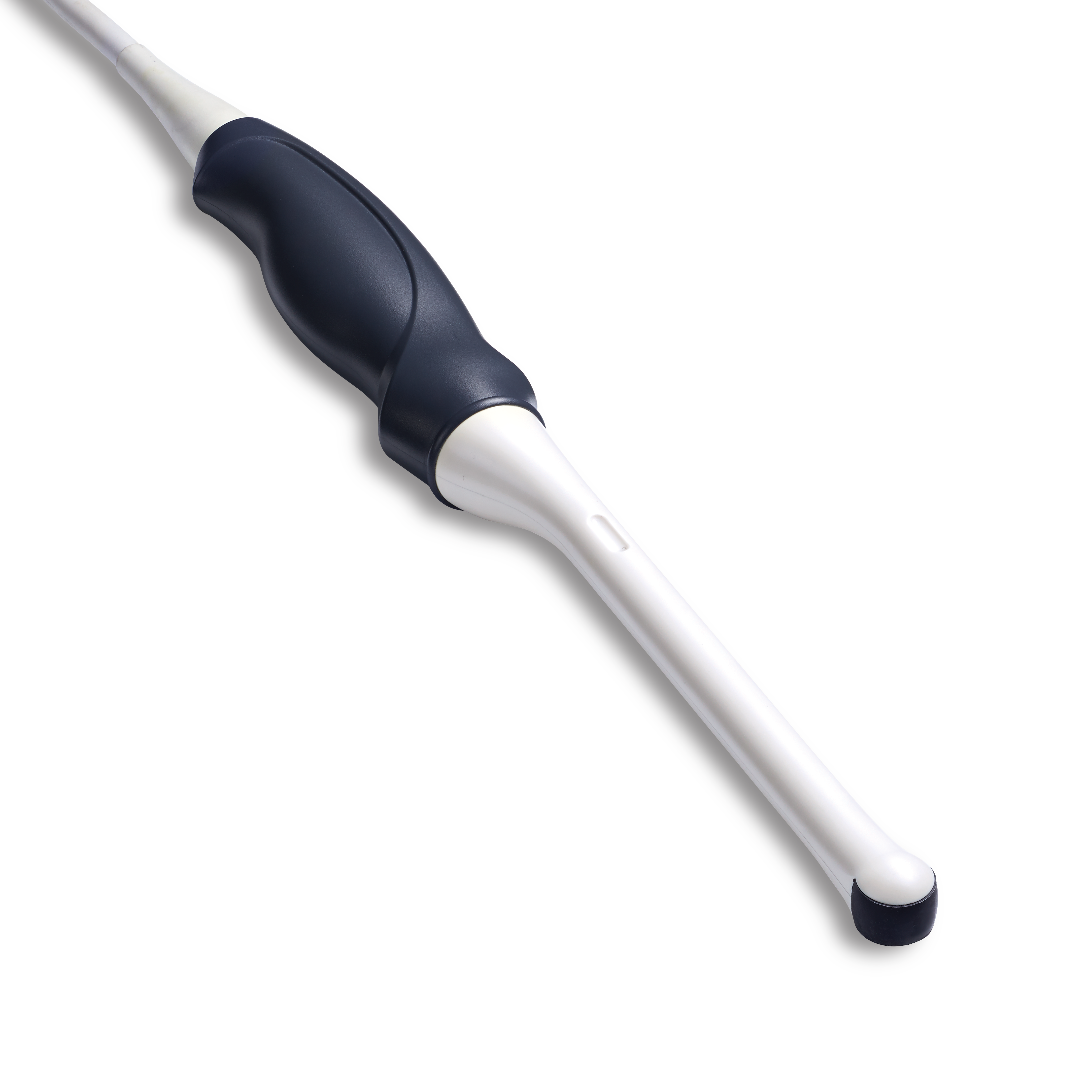
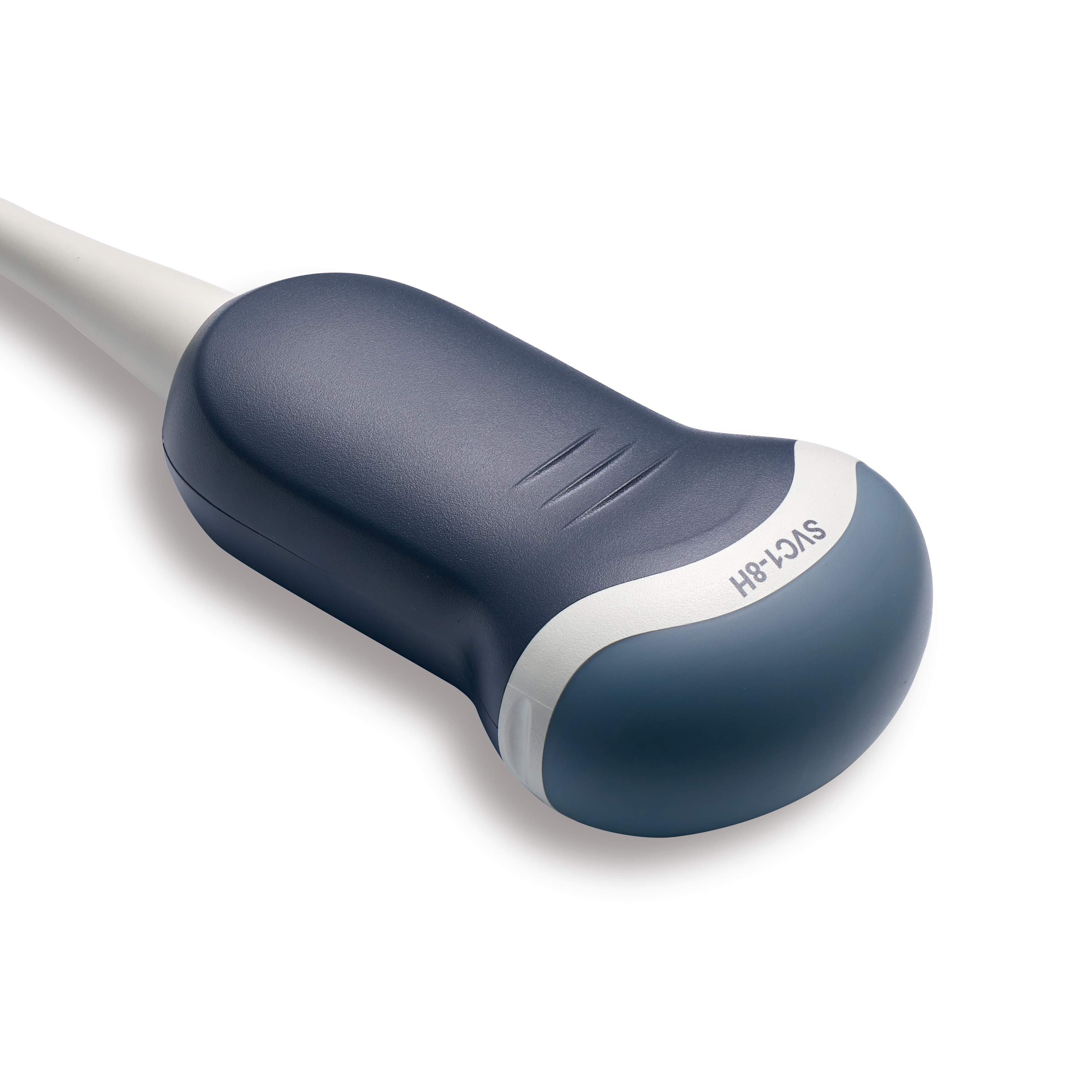
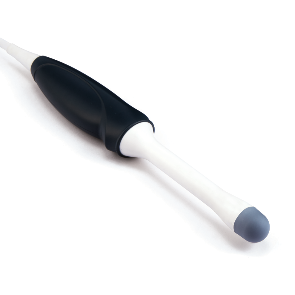
X-CUBE 60 provides healthcare professionals with a comfortable medical environment.
For confident diagnosis of a variety of diseases and conditions, optimal image performance has been realized
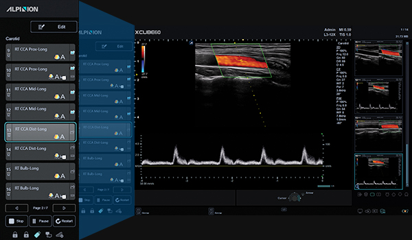
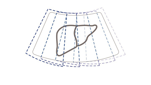
with the high-resolution image platform X+ Architecture. X-CUBE 60 is designed to improve work efficiency
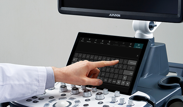
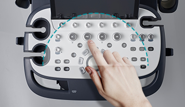
and reduce user fatigue and risk of injury. Various auto measurement tools designed for seamless workflow
quickly obtain results and the larger touch screen and newly customized control panel enhance the diagnosis comfort.
In addition, X-CUBE 60’s compact design allows for effective use of constricted scanning space.
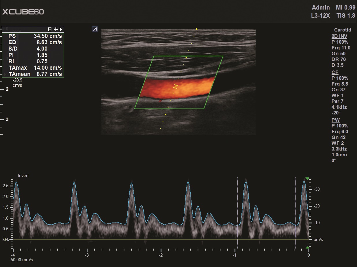
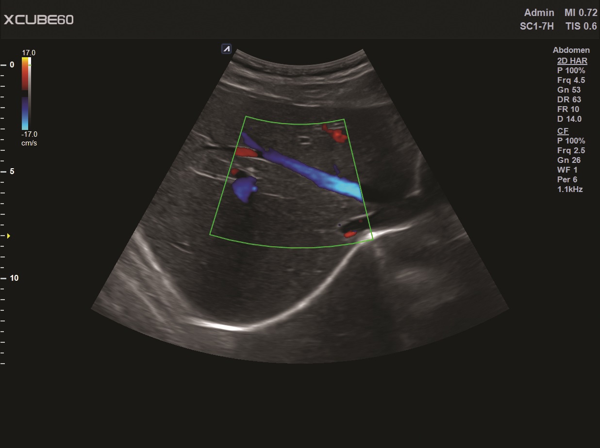
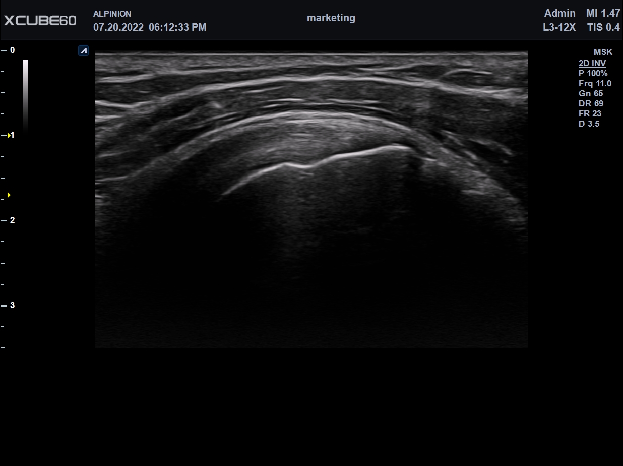
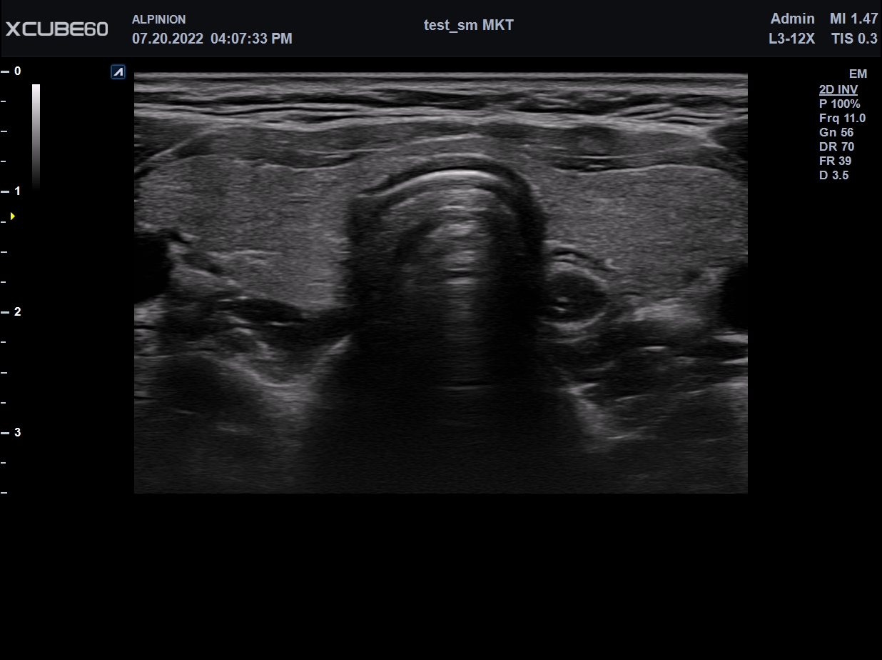
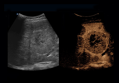
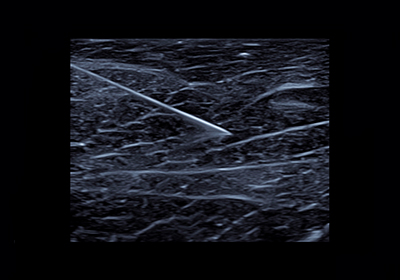
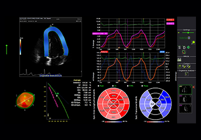
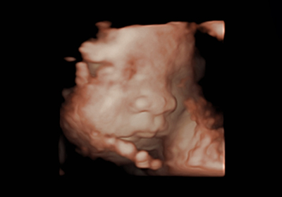
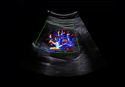
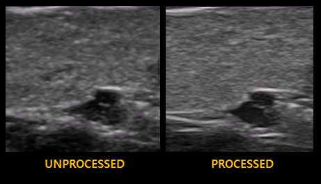
Alpinion provides professional tools for all applications such as internal medicine, OBGYN, and musculoskeletal.
This technology helps healthcare professionals achieve accurate decision and smoothly communicate with patients.
X+ MicroView is the vascular imaging mode which displays micro blood flow. Users can observe the low speed blood flow of tiny blood vessel. This technology allows for accurate diagnosis by showing a low-velocity blood flow that has not been seen in the Color Doppler at a high frame rate.
This is a function to diagnose patients using various angiographic patterns that appear while a contrast medium, administered intravenously, diffuses in blood vessels and organ tissue. CEUS has many advantages in various clinical indications for liver disease.
Using Beam Steering technology, this feature is useful in showing the shape and orientation of the needle. The needle can be viewed more clearly by adjusting the beam angle in three steps.
As a non-invasive examination method that evaluates myocardial function more objectively, it allows medical practitioners to confirm quantified data by tracking the speckle of 2D cardiac images and digitizing the movement of each myocardial segment.
Improved volume rendering technology allows for free movement of light direction and various color maps. Realistic volume image improves anatomical understanding of the fetus, allowing accurate and quick diagnosis. It also helps to form an attachment relationship between the fetus and the parent.
It is an special visualization impact technology of blood flow. It provides the intuitive understanding of vessel structure and boundaries by images of blood vessels that look like 3D on a 2D gray scale image. It may provide additional spatial information in CF, PDI or X+ MicroView.
X+ Crystal Signature™ convex transducer(1~7MHz)
Abdomen, EM, Gynecology, Obstetrics, Pediatric, Urology
X+ Crystal Signature™ Microconvex(2~11MHz)
Abdomen, Pediatric, OB/GYN, Urology, EM
High density linear transducer(8-17MHz)
Breast, EM, MSK, Vascular, Small Parts
Wideband Ultra High Freq. Linear (10~25 MHz)
Vascular, MSK, Small Parts, EM
Extreme-high density linear transducer (3-12MHz)
Breast, EM, MSK, Vascular, Small Parts
High density linear transducers (3-15 MHz)
MSK, Vascular, Small Parts, Breast, EM
X+ Crystal Signature™
Extreme-high density linear transducer (3-19MHz)
Abdomen, Pediatric, Gynecology, Obstetrics,
EM, MSK, Vascular, Small Parts, Breast, TCD
Single crystal phased array transducer (1-5MHz)
Abdomen, Cardiac, EM, TCD
X+ Crystal Signature endocavity transducer(2~11MHz)
Gynecology, Obstetrics, Urology, EM
X+ Crystal Signature endocavity transducer(2~11 MHz)
Gynecology, Obstetrics, Urology, EM
Single Crystal Volume Convex
Gynecology, Obstetrics, Abdomen, Pediatric, Urology, EM
High density volume endocavity transducer (3-10MHz)
GYN, OB, Urology, EM
Pencil type transducer (5.0MHz)
Cardiac
Pencil type transducer (2.0MHz)
Cardiac
Pencil type transducer (8.0MHz)
Cardiac