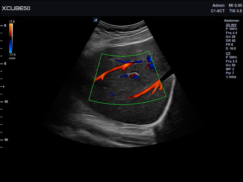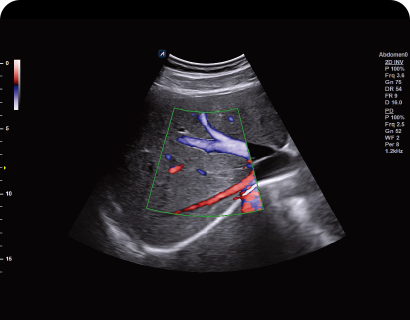
C1-6CT
C-Architecture (PowerView™) Convex transducer (1-6MHz)
Application:
Abdomen, EM, Gynecology, Obstetrics
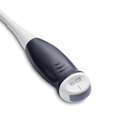
SC2-11H
X+ Crystal Signature™ Microconvex(2~11MHz)
Application:
Abdomen, Pediatric, OB/GYN, Urology, EM
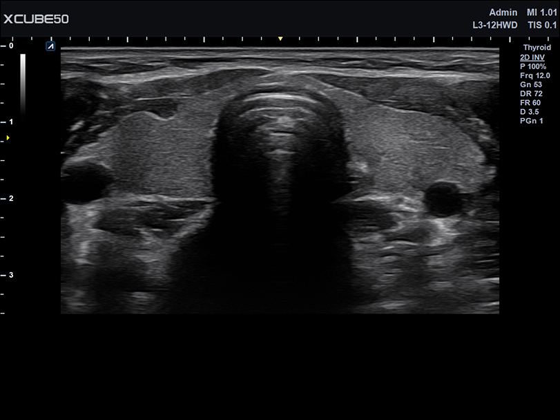
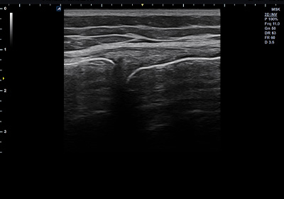
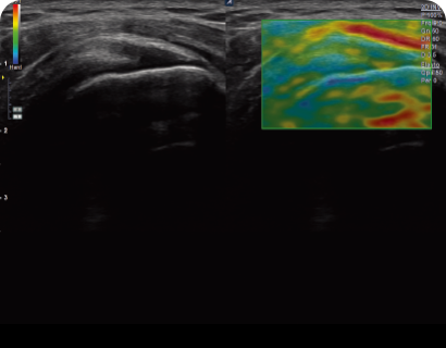
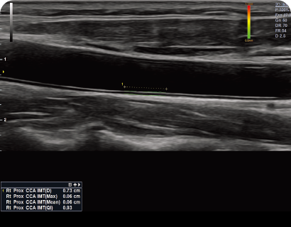
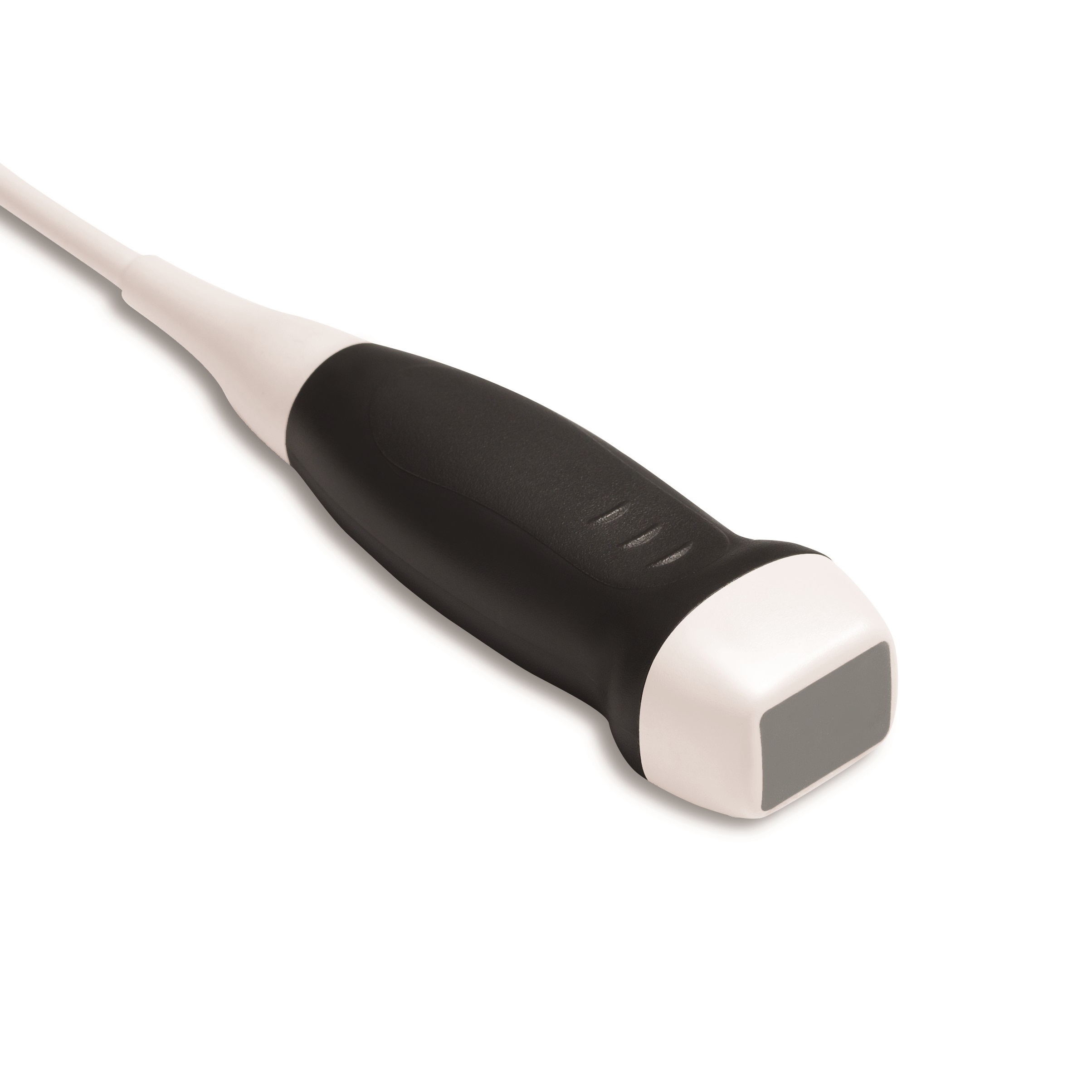
L3-12HWD
High density wide footprint linear transducer (3-12MHz)
Application:
Breast, EM, MSK, Vascular, Small Parts, Appendix
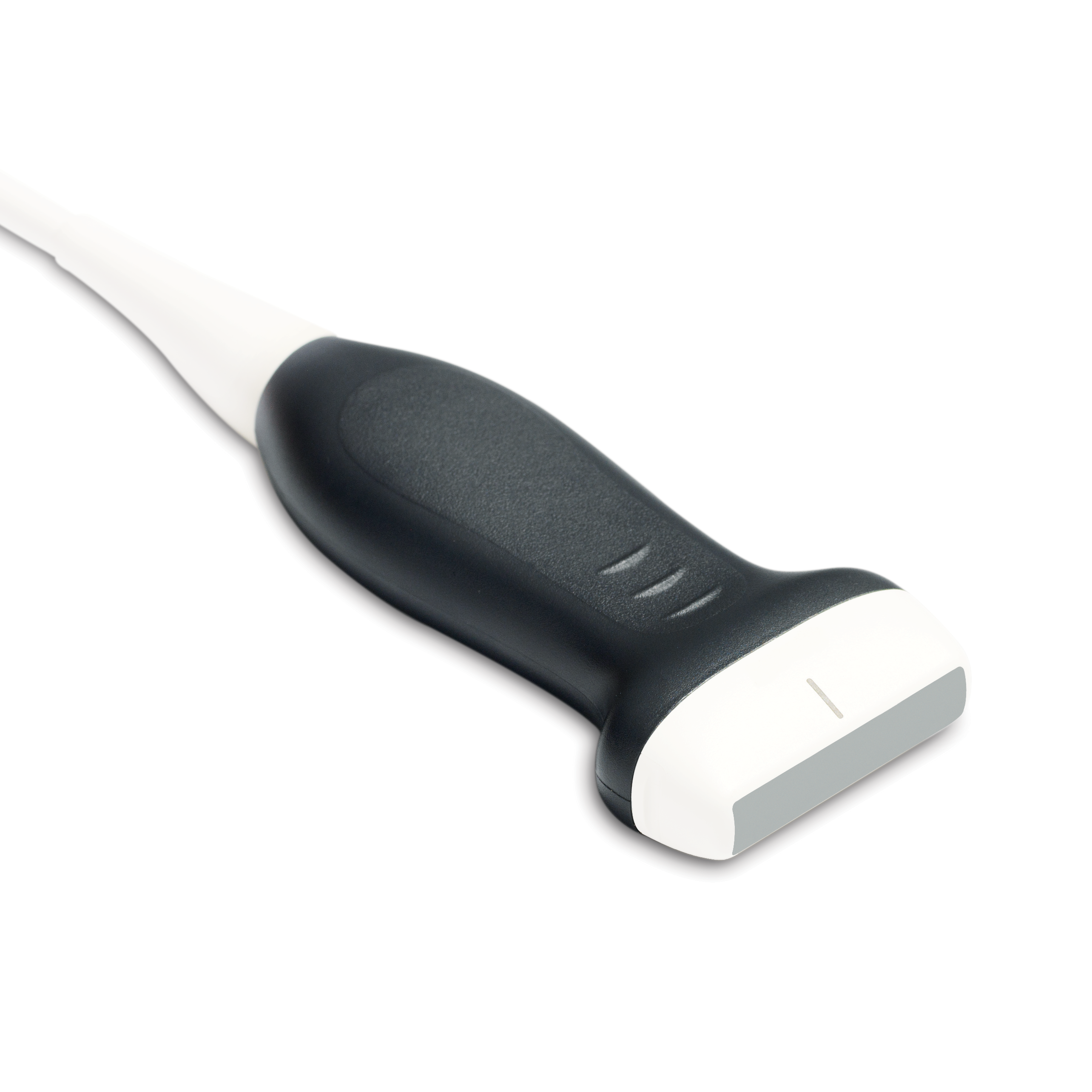
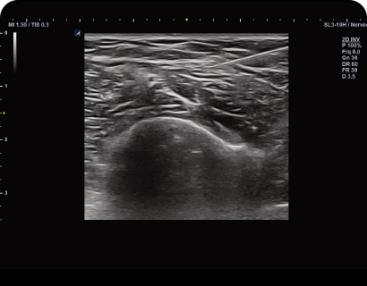
L3-8H
High density low frequency linear transducer (3-8MHz)
Application:
Breast, EM, MSK, Vascular, Small Parts
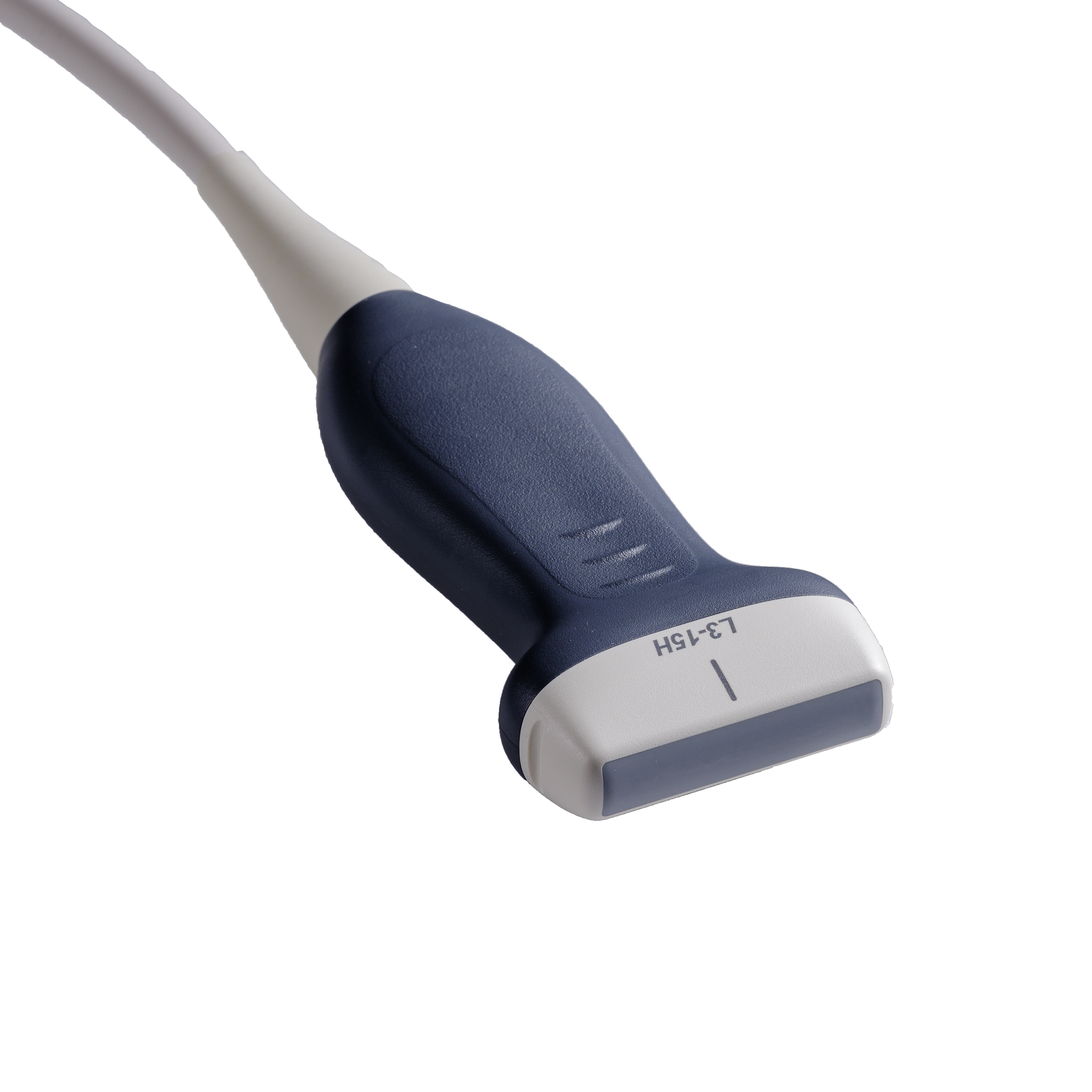
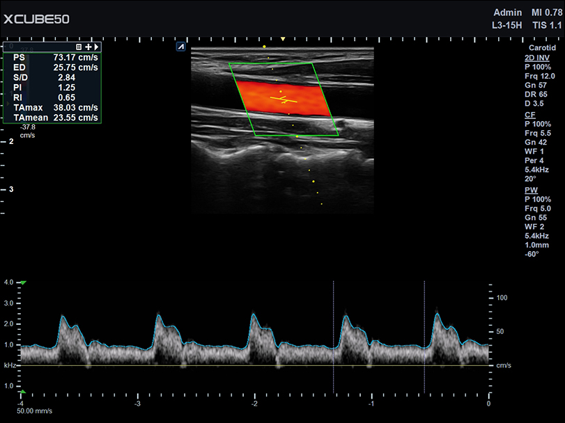
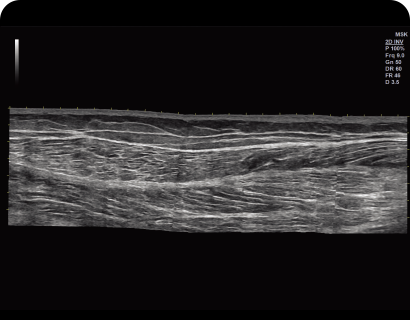
L3-15H
High density linear transducers (3-15 MHz)
Application:
MSK, Vascular, Small Parts, Breast, EM
P1-5CT
Single crystal phased array transducer (1-5MHz)
Application:
Abdomen, Cardiac, EM, TCD
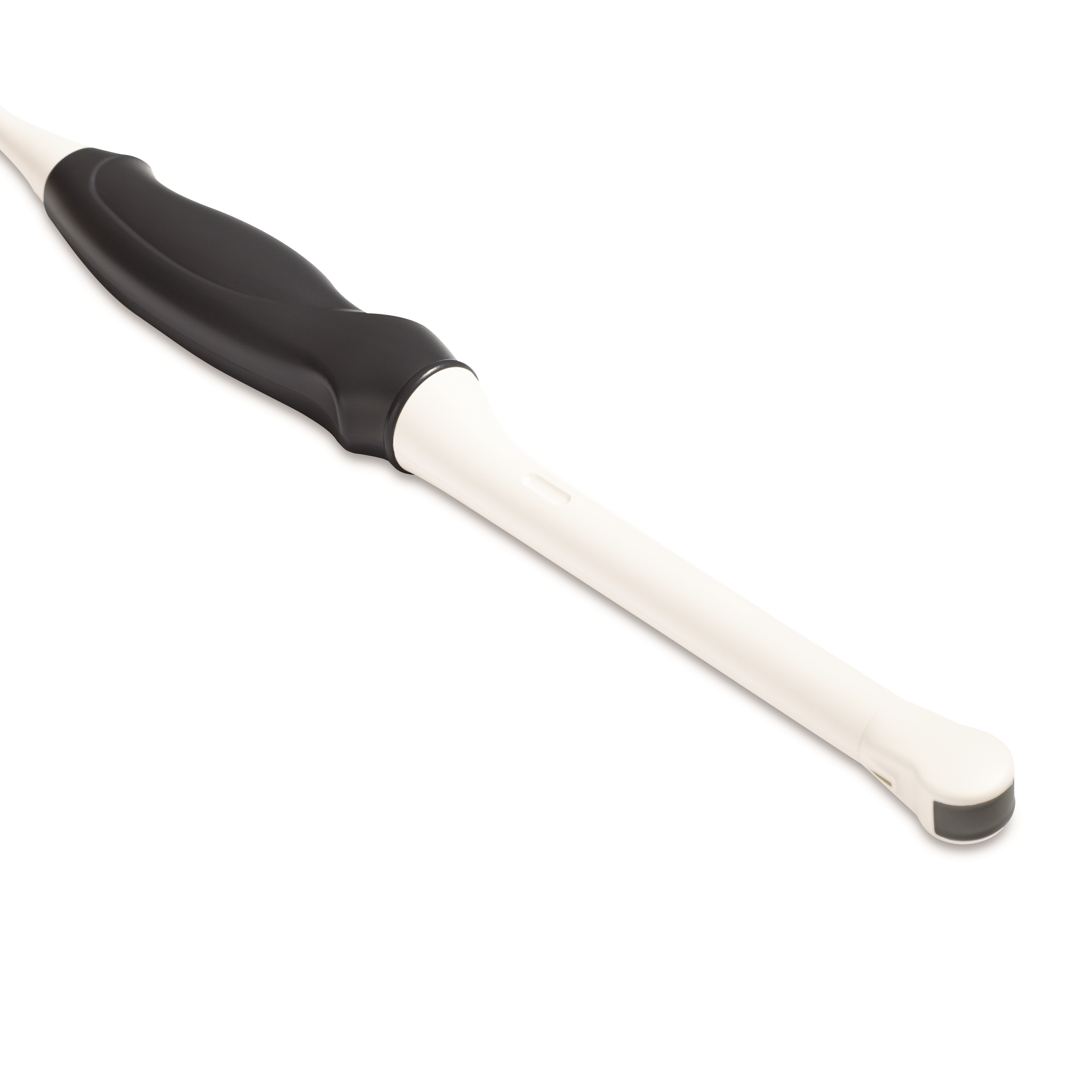
EV3-10T
Endocavity transducer (3-10MHz)
Application:
GYN, OB, Fetal Echo, Urology, EM
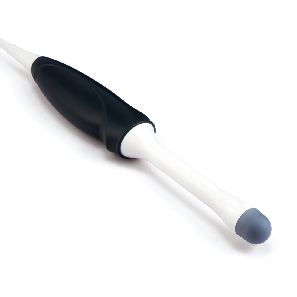
EC3-10T
Endocavity transducer (3-10MHz)
Application:
GYN, OB, Fetal Echo, Urology, EM
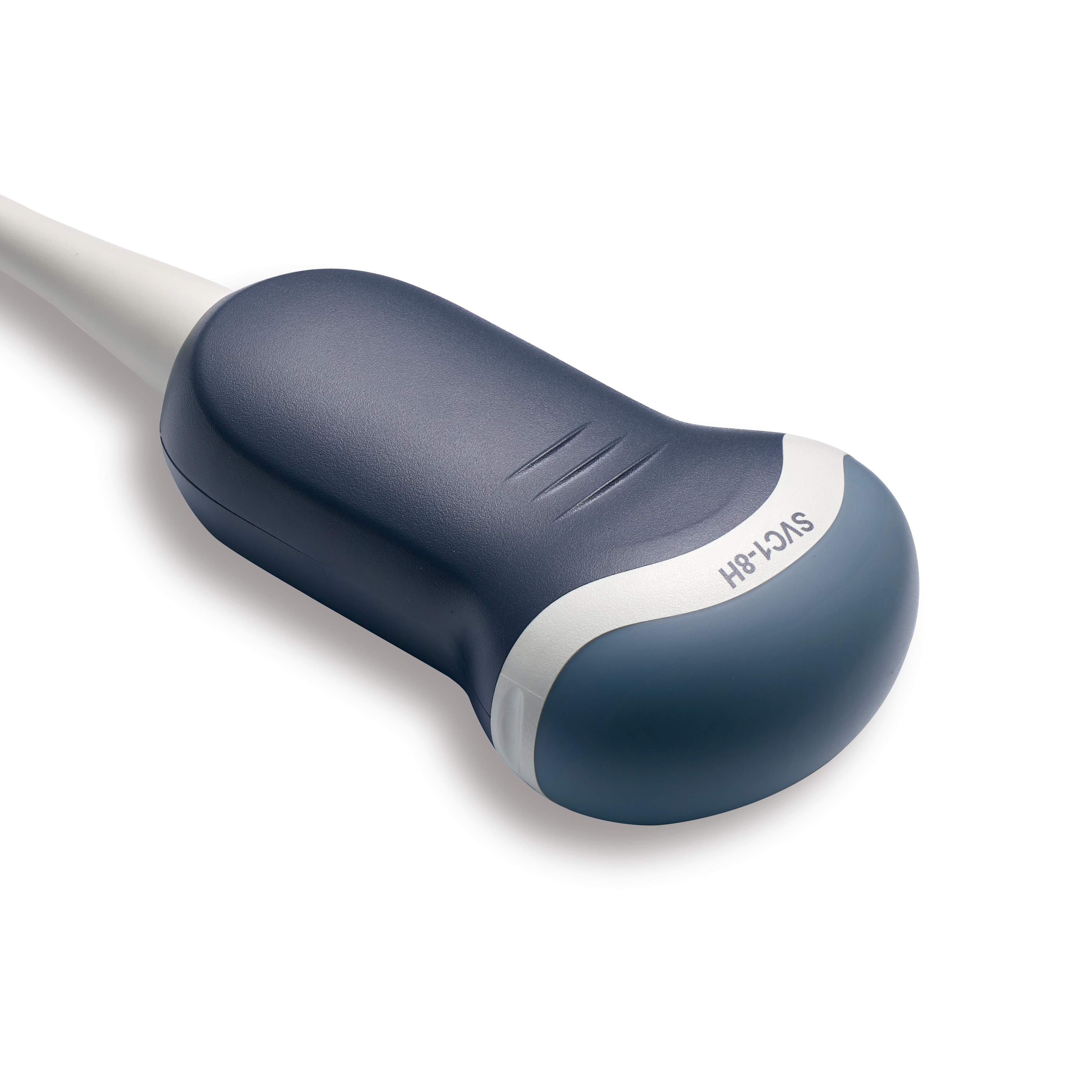
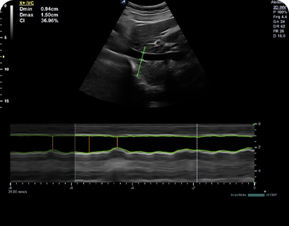
SVC1-8H
Single Crystal Volume Convex
Application:
Gynecology, Obstetrics, Abdomen, Pediatric, Urology, EM
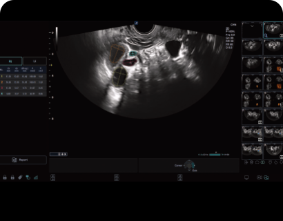
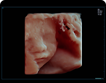
VE3-10H(NEW)
Volume Endocavity
Application:
Gynecology, Obstetrics, Urology, EM
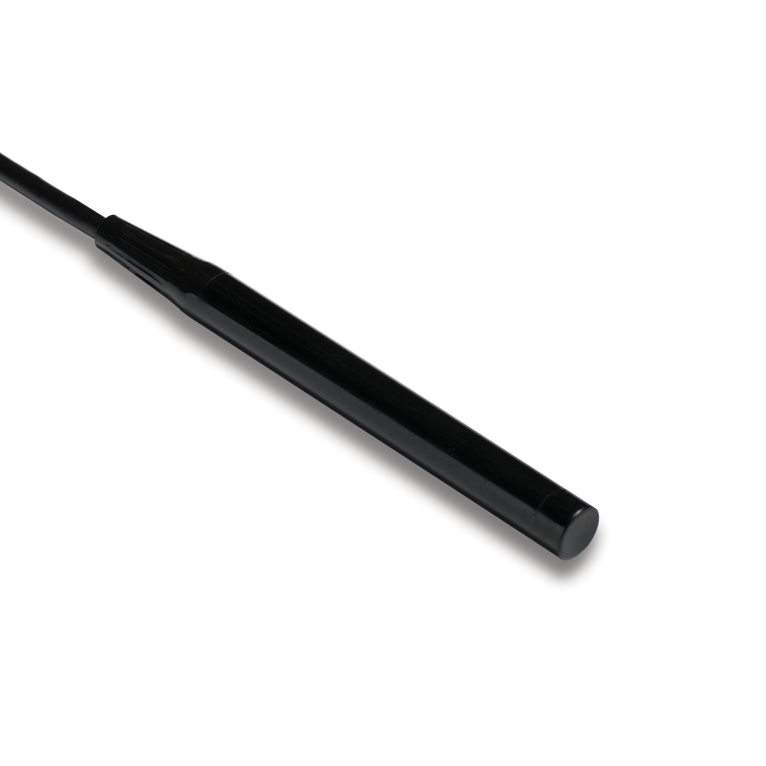
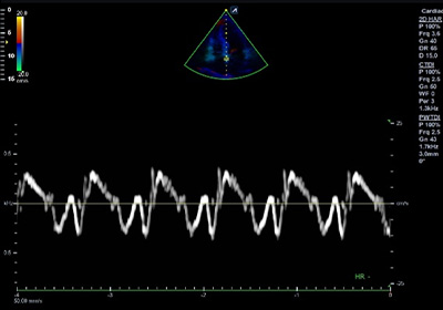
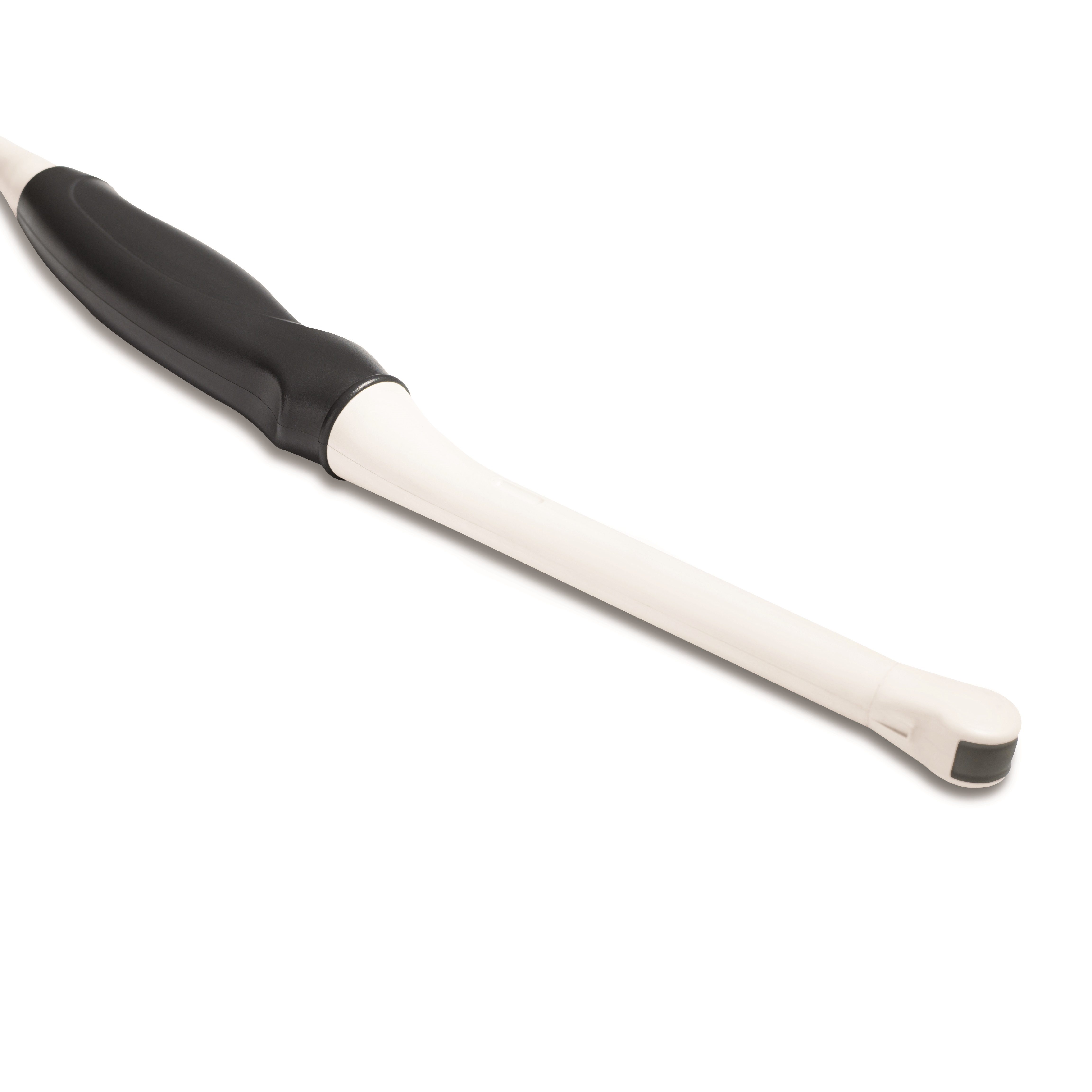
CW5.0
Pencil type transducer (5.0MHz)
Application:
Cardiac

CW2.0
Pencil type transducer (2.0MHz)
Application:
Cardiac

CW8.0
Pencil type transducer (8.0MHz)
Application:
Cardiac