
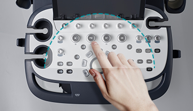

X+ Assistant
Keystrokes have been reduced by more than 50% compared to conventional methods, thereby reducing examination time. Optimal scanning protocols are registered based on application -specific guidelines, allowing users to personalize and optimize protocols.

Power Preset
Users can load a system preset saved in advance with a single button click. By using these quick and easy presets, users can shorten the imaging set up time.

X+ Auto Biometry
When measuring fetal EFW, Auto Biometry detects fetal HC, BPD, FL, AC, and Humerus, and automatically measures their lengths.

X+ Compare
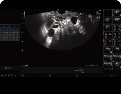
Users can import previously acquired videos from a PACS or hard disk and compare them with the current videos in real-time scan, which is a key feature to optimize patient care. * X+Compare supports ultrasound studies only

Xpeed™
Simply press the Xpeed™ button once to quickly optimize images in 2D Mode and Spectrum Doppler Mode. Detect, predict, and adjust the Dynamic range level in real-time.

USB Real-time recording
USB real-time recording simplifies data transfer by enabling users to record ultrasound scan images directly onto USB memory in real time. Videos are captured in high-definition and quickly stored in the system.
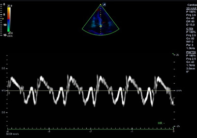
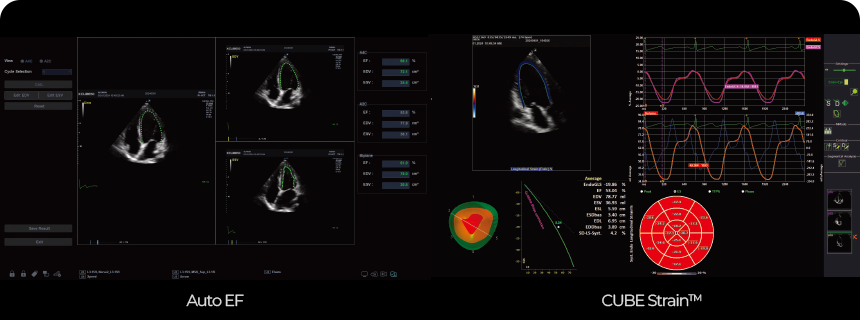
Echo Package : Auto EF, CUBE Strain™
It is a non-invasive test method that automatically analyzes the end-diastolic volume (EDV), end-systolic volume (ESV), and ejection fraction (EF), providing a more objective evaluation of myocardial function. Additionally, it quantifies data by tracking speckles in 2D heart images to assess myocardial movement.
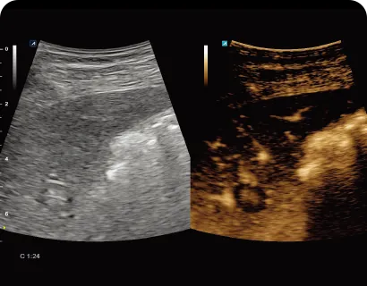
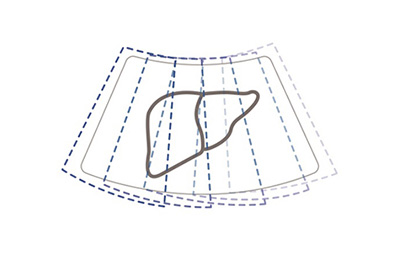
Contrast Enhancement Ultrasound (CEUS)
This function diagnoses patients using various angiographic patterns that appear while a contrast medium is administered intravenously, diffusing in blood vessels and organ tissue. It is useful for diagnosing intrahepatic masses, examining liver tissue, and assessing the severity of liver fibrosis and portal hypertension.
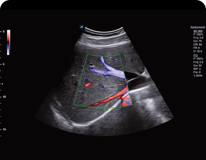
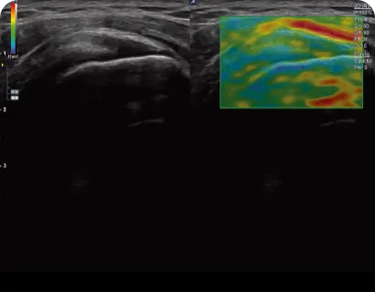
Directional PD Mode (DPDI)
Power Doppler technology displays blood flow direction information with higher sensitivity than Color Doppler.
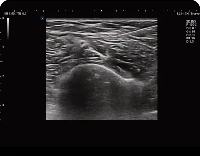
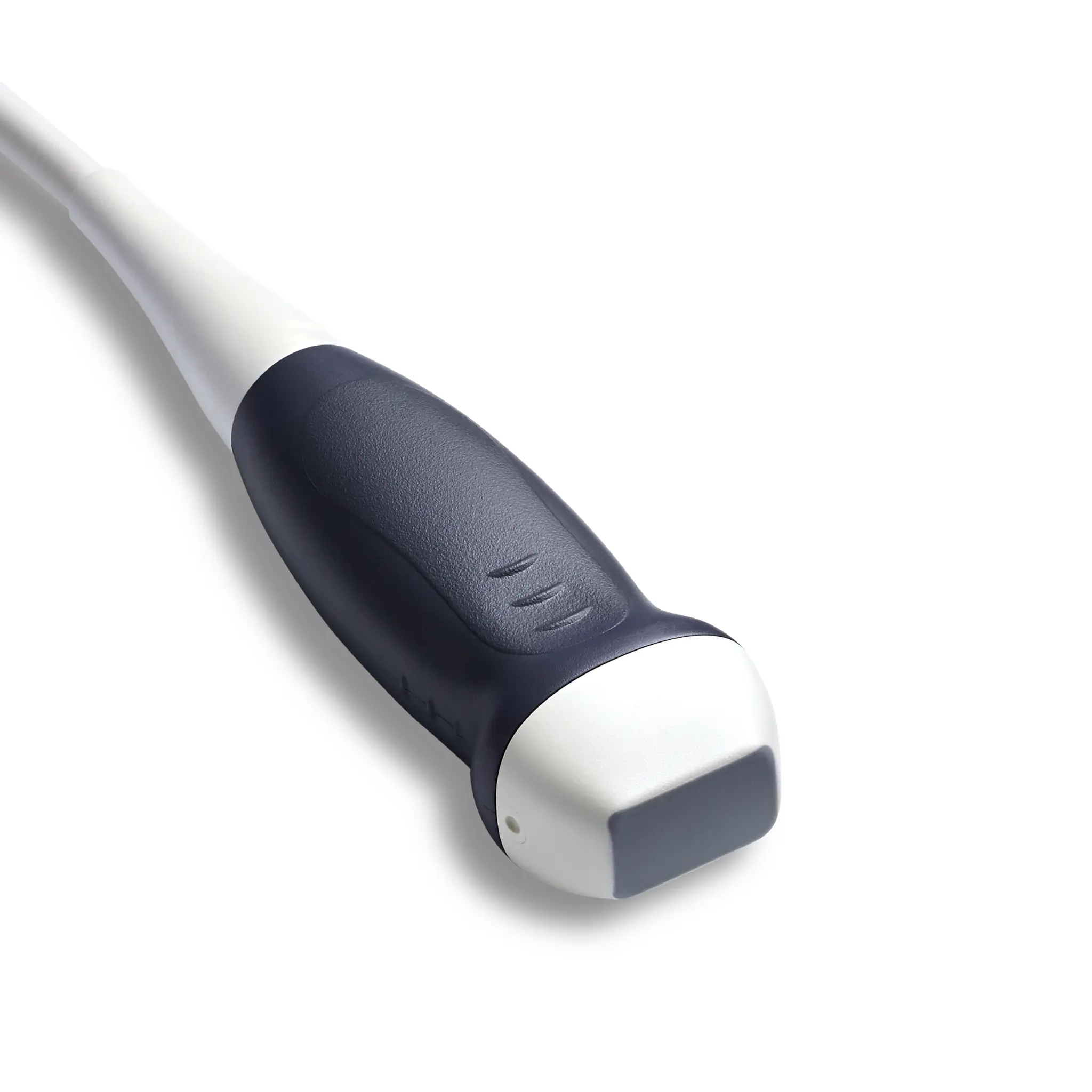
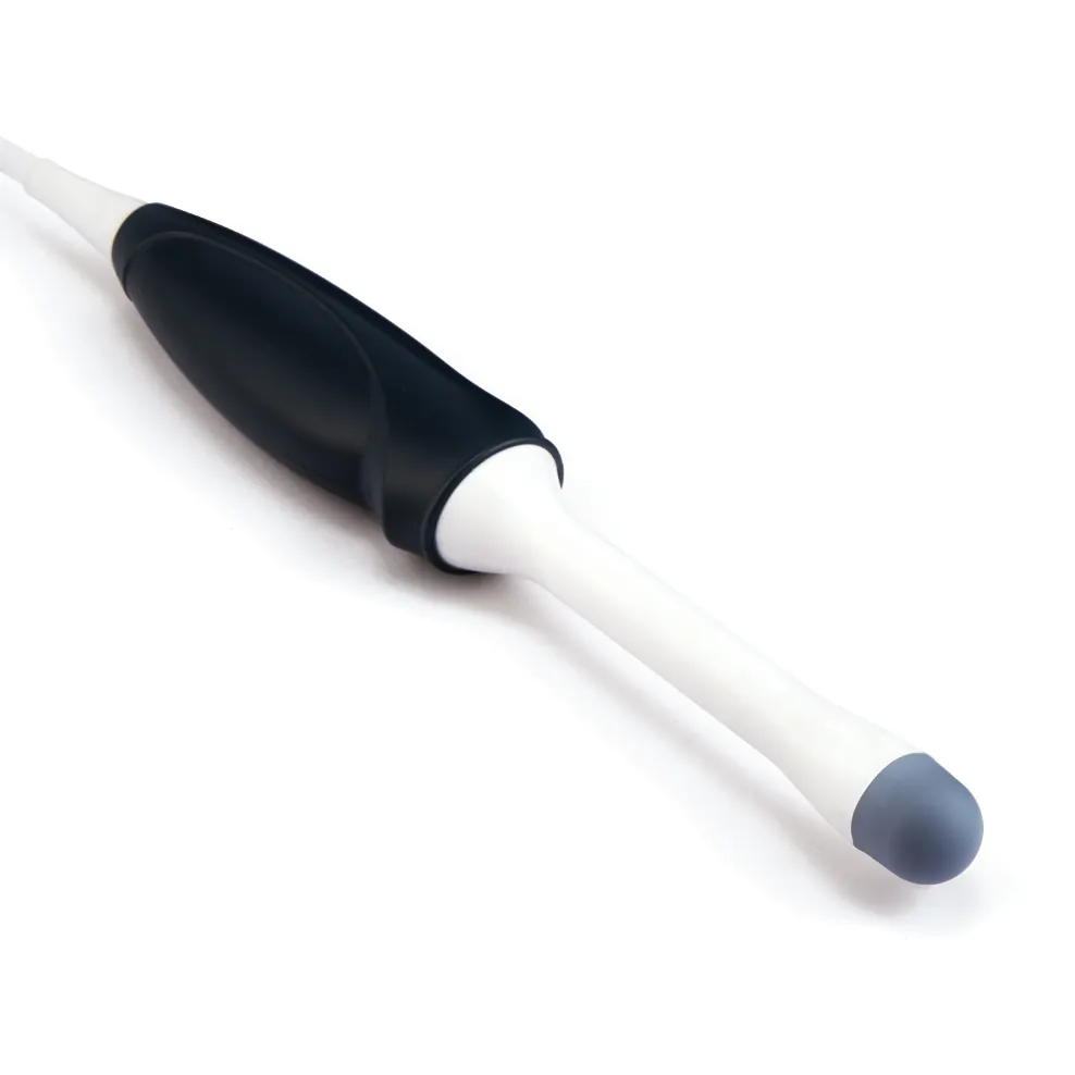
Needle Vision™ Plus
This function is useful for clearly displaying the shape and direction of the needle during invasive testing, employing beam steering technology.
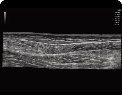
Panoramic
Panoramic imaging enables observation of extremely wide horizontal images.
Strain Elastography
Elastic ultrasound imaging is an ultrasound imaging technique that revels relative elasticity of tissues against external pressure. It provides additional pathological information, helping to reduce unnecessary biopsies.
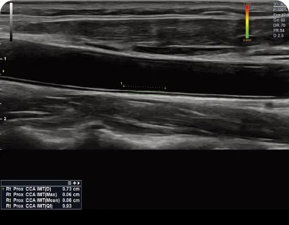
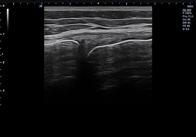
Auto IMT
The thickness of the carotid artery intima-media can be accurately and quickly measured down to the millimeter unit, regardless of the user's skill level.
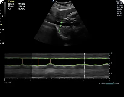
Auto IVC
This function can automatically distinguish the inferior vena cava (IVC) from surrounding blood vessels and tissues and measure changes in diameter, which make it convenient for evaluating right atrium function.
Auto Follicle
To measure superovulation, this technology counts the number of follicles and measures the volume automatically.
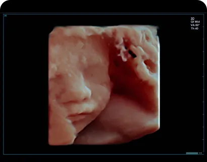
Live HQ™
The improved volume rendering technology allows for the free movement of light direction and supports various color maps.